An instrument capable of imaging cellular details previously invisible even to modern microscopes has arrived at our Imaging Facility.
The new super-resolution microscope combines three advanced technologies and offers sharper images as well as the ability to observe how individual molecules interact within cells. Our institute thus gains a tool that will expand the possibilities to investigate processes important for plant growth and behavior. The equipment will also be available to scientists from other institutions, who can use it for their projects.
The Imaging Facility of the Institute of Experimental Botany of the Czech Academy of Sciences (IEB CAS) is equipped with several state-of-the-art confocal microscopes, which are the standard in today‘s biological research. They image samples with incomparably higher quality than standard school microscopes. However, technological development is advancing rapidly, and new microscopic techniques are emerging that transcend the limits of classical optics; this is precisely why the institute purchased a super-resolution microscope equipped with the most modern technologies this year.
The Mirava Polyscope instrument was supplied by the German company Abberior Instruments. “We are the first institution in Central Europe where this model has been installed. Furthermore, the system was custom-tailored for plant microscopy. We are gradually discovering its capabilities and are thrilled with the quality of the data obtained,” says Kateřina Malínská, head of the Imaging Facility at the IEB CAS.
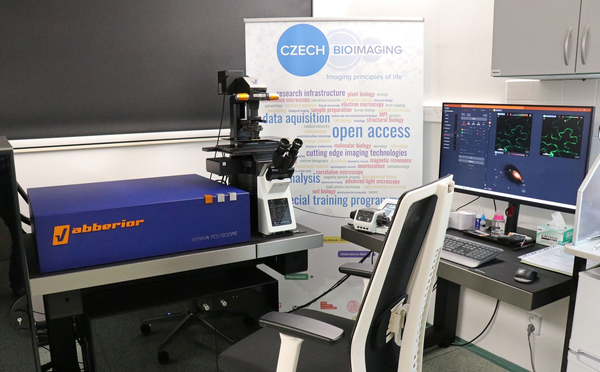
The new Mirava Polyscope microscope at the Institute of Experimental Botany of the Czech Academy of Sciences.
Seeing More and Better
The microscope offers three advanced imaging technologies that utilize a phenomenon called fluorescence—that is, the ability of substances to emit light of a different color after being illuminated by light of a specific wavelength. For example, banknotes have fluorescent security features that glow in various colors of visible light under an ultraviolet lamp. In biology, fluorescence can be used to “label” specific intracellular structures or protein molecules.
The first technology is super-resolution microscopy, which can image and resolve structures ten times smaller than conventional microscopy. The groundbreaking discovery of super-resolution was awarded the Nobel Prize in 2014, and one of its laureates, Stefan Hell, founded the company Abberior Instruments with his colleagues.
The second technology is FLIM (Fluorescence Lifetime Imaging Microscopy), an advanced technique in fluorescence microscopy that measures not only the color and intensity of fluorescence like a standard confocal microscope, but also how long it takes for the fluorescence to fade after illumination. This “decay time” is characteristic of each substance and makes it surprisingly easy to distinguish them.
“In plants, for example, the fluorescence of the green pigment chlorophyll often interferes. FLIM can separate it from the fluorescence of the structure or molecule we want to investigate, which significantly improves image quality,” explains Matěj Drs from the IEB CAS Imaging Facility. The method also enables to observe interactions between proteins, because the decay time changes when protein molecules come into close proximity.
The third technology is adaptive optics, originally developed for astronomical telescopes. In microscopes, it continuously adjusts the optical path according to the nature of the sample to ensure the highest possible image quality. This makes it possible to observe even deeper tissue layers, such as vascular bundles inside living roots.
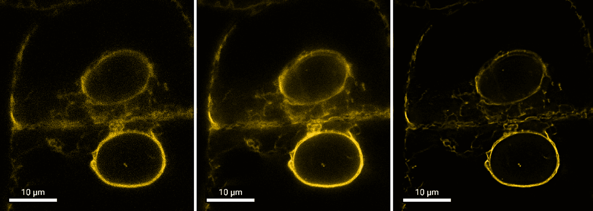
Plant cells with cell nuclei (two circular structures). The image on the left was taken using conventional confocal microscopy, the one in the middle using super-resolution microscopy, and the one on the right using a combination of super-resolution microscopy and FLIM. This combination provides such a high resolution that the two membranes forming the nuclear envelope can be clearly distinguished from one another. Photo by Jana Krtková, sample by Lenka Helusová, both from the IEB CAS.
Open to other research teams
“Individual technologies can be used separately, but they can also be combined, which further expands the microscope’s capabilities. Following initial testing and its debut at the fall microscopy course, Mirava is now entering full operation, and the first research projects are beginning. Its range is very diverse, from the study of moss cells and cell wall structures in high resolution, through protein interactions in pollen, to research on proteins that transport plant hormones,” explains Kateřina Malínská.
External institutions have also already expressed interest in the new instrument. The IEB CAS is a member of the national research infrastructure Czech-BioImaging, which brings together institutions focused on imaging in biological and medical research.
The purchase of the instrument was funded by the Ministry of Education, Youth, and Sports through the Jan Amos Komenský Operational Program for research infrastructure. Part of the costs was also covered by the laboratories of IEB CAS.
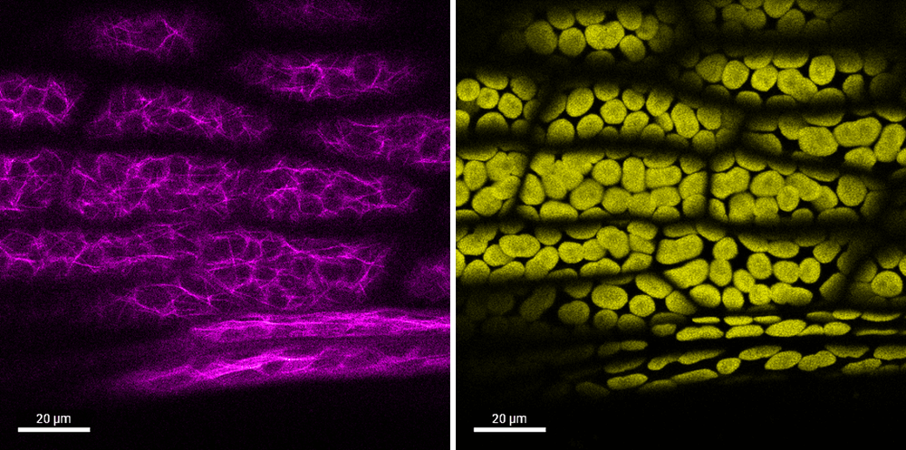
FLIM technology allows for the differentiation of fluorescence originating from various sources. Both images show the same sample—leaf cells of the spreading earthmoss (Physcomitrium patens). On the left, you can see fluorescently labeled protein filaments called microtubules, which are important e.g. for intracellular transport or cell division. The image on the right shows the fluorescence of the green pigment chlorophyll. Photo by Matěj Drs, sample by Chandran Kalathodi Laiju, both from the IEB CAS.
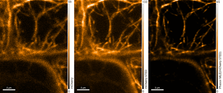
A highly detailed view of actin protein filaments, which control the movements of various intracellular structures. The image on the left was taken using standard confocal microscopy. In the middle is a higher-quality image obtained using super-resolution microscopy. Resolution improves further when super-resolution microscopy is combined with the FLIM technique (right). Photo: Kateřina Malínská, IEB CAS.